Osteochondral Lesion of the Talus (Ankle)
Osteochondral Lesions of the Talus (Ankle)
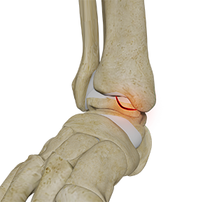
The tibia and the fibula bones of the lower leg join with the talus bone to form the ankle joint. The talus bone is an important bone located between the tibia and fibula and the heel bone (calcaneus). OCL or OCD is the damage to the cartilage and the talus bone of the ankle joint. Usually, the inner or the medial portion of the ankle is affected.
Osteochondral (OCL) injuries of the ankle (Talus and Tibia) as well as other joints including the 1st MTP, subtalar and navicular are common. The division has more experience in this condition than any other facility in the US with over 100 publications reporting on outcomes. The division surgeons were pioneers in using concentrated bone marrow aspirate in OCL of the ankle and continues to be at the forefront of stem cell therpy, autologous osteochondral transplant (OATS) and other cartilage sparing techniques.
Where can I have Osteochondral Lesions?
- Talus (talar dome: medial and lateral)
- Tibia
- 1st MTP
- Subtalar Joint
- Navicular
- Femur (Knee)
Causes of OCL of the Talus (Ankle)
OCL may be genetic or may be caused due to trauma, stress fractures in sports, severe sprain, local osteonecrosis, etc.
Symptoms of OCL of the Talus (Ankle)
OCL lesions are sometimes asymptomatic. Large lesions are associated with symptoms such as localized ankle pain and discomfort which worsens while walking or running, a clicking or popping sound, swelling, tenderness, weakness of the foot, etc.
Diagnosis of OCL of the Talus (Ankle)
Osteochondral lesions are diagnosed by a physical examination, X-ray, and CT and MRI scans. Plain X-ray images can reveal other fractures, bone spurs, and narrowing of the joint. A CT scan helps identify any bony fragments but is not very helpful to visualize bone edema or cartilage defects. MRI is the best imaging modality, which helps to visualize the cartilage and bone lesions as well as bone edema.
Treatment of OCL of the Talus (Ankle)
Non-surgical Method
Small lesions can be treated non-surgically involving the use of a cast, physical therapy, pain medications, strengthening exercises, etc.
Arthroscopy
An arthroscope is a narrow tube with a tiny video camera on one end. The structures inside the ankle are visible to your surgeon on a monitor in the operating room. Your doctor treats OCL by removing the damaged tissue followed by microfracture, which enables natural healing of the damaged bone and cartilage.
OATS Procedure
Large lesions are treated by the OATS procedure. OATS of the ankle is a surgical procedure to treat Osteochondral Lesions of the Talus (OCL) or Osteochondritis Dissecans (OCD). It involves the transfer of healthy cartilage to replace the damaged cartilage and restore the normal function of the foot. The cartilage can be taken from your ankle joint (autograft) for smaller defects. An allograft (graft from a donor) is considered for large defects. During an OATS procedure, multiple, tiny plugs of healthy bone and cartilage are transferred in a pattern that resembles a mosaic, hence, the procedure is also known as mosaicplasty.
Prevention of OCL of the Ankle
OCL of the ankle can be prevented by:
- Learning proper techniques in sports
- Minimizing overuse activities
- Practicing strength training exercises
Dr. John G. Kennedy, MD | Osteochondral Defects
Osteochondral Lesions in the Ankle by Dr. John G. Kennedy
Dr. Kennedy’s Publications links:
- Osteochondral Defects of the Talus: Current Management Dilemmas
- Knee-to-Talus Donor-Site Morbidity Following Autologous Osteochondral Transplantation: A Meta-Analysis with Best-case and Worst-case Analysis
- Lesion Size Measured on MRI Does Not Accurately Reflect Arthroscopic Measurement in Talar Osteochondral Lesions
- Biologic Adjuvants for the Management of Osteochondral Lesions of the Talus
- Post-treatment Follow-up, Imaging, and Outcome Scores: Proceedings of the International Consensus Meeting on Cartilage Repair of the Ankle
- Conservative Management and Biological Treatment Strategies: Proceedings of the International Consensus Meeting on Cartilage Repair of the Ankle
- Scaffold-Based Therapies: Proceedings of the International Consensus Meeting on Cartilage Repair of the Ankle
- Incidence of Coexisting Talar and Tibial Osteochondral Lesions Correlates With Patient Age and Lesion Location
- Effect of the Containment Type on Clinical Outcomes in Osteochondral Lesions of the Talus Treated With Autologous Osteochondral Transplantation
- High reported rate of return to play following bone marrow stimulation for osteochondral lesions of the talus
- Good clinical and functional outcomes at mid-term following autologous osteochondral transplantation for osteochondral lesions of the talus
- Systematic review of bone marrow stimulation for osteochondral lesion of talus - evaluation for level and quality of clinical studies
- Platelet-Rich Plasma and Hyaluronic Acid Are Not Synergistic When Used as Biological Adjuncts with Autologous Osteochondral Transplantation
- Subchondral Bone Degradation After Microfracture for Osteochondral Lesions of the Talus: An MRI Analysis
- The Subchondral Bone Is Affected by Bone Marrow Stimulation: A Systematic Review of Preclinical Animal Studies
- Osteochondral lesions of the talus in the athlete: up to date review
- Current management of talar osteochondral lesions
- Operative Treatment for Osteochondral Lesions of the Talus: Biologics and Scaffold-Based Therapy
- Platelet-Rich Plasma and Concentrated Bone Marrow Aspirate in Surgical Treatment for Osteochondral Lesions of the Talus
- Diagnosis and treatment of osteochondral lesions of the ankle: current concepts
- Magnetic Resonance Imaging Evidence of Postoperative Cyst Formation Does Not Appear to Affect Clinical Outcomes After Autologous Osteochondral Transplantation of the Talus
- Clinical and MRI Donor Site Outcomes Following Autologous Osteochondral Transplantation for Talar Osteochondral Lesions
- Autologous Osteochondral Transplantation for Osteochondral Lesions of the Talus
- Arthroscopic Bone Marrow Stimulation and Concentrated Bone Marrow Aspirate for Osteochondral Lesions of the Talus: A Case-Control Study of Functional and Magnetic Resonance Observation of Cartilage Repair Tissue Outcomes
- Autologous osteochondral transplantation for osteochondral lesions of the talus in an athletic population
- Anterolateral tibial osteotomy for accessing osteochondral lesions of the talus in autologous osteochondral transplantation: functional and t2 MRI analysis
- Functional and MRI outcomes after arthroscopic microfracture for treatment of osteochondral lesions of the distal tibial plafond
- Osteochondral lesions of the talus: a current concepts review and evidence-based treatment paradigm
- Talonavicular arthroscopy for osteochondral lesions: technique and case series
- Double-Plug Autologous Osteochondral Transplantation Shows Equal Functional Outcomes Compared With Single-Plug Procedures in Lesions of the Talar Dome: A Minimum 5-Year Clinical Follow-up
- Osteochondral lesions of the talus: aspects of current management
- Operative treatment of osteochondral lesions of the talus
- A systematic review on the reporting of outcome data in studies on autologous osteochondral transplantation for the treatment of osteochondral lesions of the talus
- Microfracture for osteochondral lesions of the talus: a systematic review of reporting of outcome data
- Critical defect size for osteochondral lesions of the talus: letter to the editor
- Establishing proof of concept: Platelet-rich plasma and bone marrow aspirate concentrate may improve cartilage repair following surgical treatment for osteochondral lesions of the talus
- The Treatment of Osteochondral Lesions of the Talus with Autologous Osteochondral Transplantation and Bone Marrow Aspirate Concentrate: Surgical Technique
- Current concepts in the diagnosis and treatment of osteochondral lesions of the ankle
- Arthroscopic-assisted fluoroscopic navigation for retrograde drilling of a talar osteochondral lesion
- Osteochondral lesion of the fifth metatarsal head in a triathlete
- Platelet-Rich Plasma May Improve Osteochondral Donor Site Healing in a Rabbit Model
- Platelet-rich plasma increases transforming growth factor-beta1 expression at graft-host interface following autologous osteochondral transplantation in a rabbit model
- Autologous osteochondral transplantation for osteochondral lesions of the talus: high rate of return to play in the athletic population