Osteochondral Autograft Transplantation System (OATS) of the Ankle | OATS Procedure
Dr. Kennedy has performed more OATS procedures and published more studies on them than anyone in the USA
What is OATS of the Ankle?
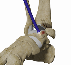
OATS of the ankle is a surgical procedure to treat Osteochondral Lesions of the Talus (OCL) or Osteochondritis Dissecans (OCD). It involves the transfer of healthy cartilage to replace the damaged cartilage and restore the normal function of the foot. The cartilage can be taken from your ankle joint (autograft) for smaller defects. An allograft (graft from a donor) is considered for large defects. During an OATS procedure, multiple, tiny plugs of healthy bone and cartilage are transferred and laid in a mosaic pattern, hence, the procedure is also known as mosaicplasty.
OATS Ankle Procedure
The surgery is performed under general anesthesia. It involves the following steps:
- An incision is made at the ankle joint. A few ligaments may be incised to expose the joint.
- Your surgeon performs debridement of the chondral surface to remove damaged cartilage. Some damaged bone may also be removed. Care is taken to prevent damage to healthy cartilage.
- An autograft is procured from a non-articulating portion of your knee and is inserted at the damaged site. Great cone is needed to ensure perfect alignment.
- The tibial osteotomy is fixed with titanium screws to allow post-operative MRI.
- Stem cells are used to augment healing.
What are Osteochondral Lesions (OCL)?
OCL’s are defects of the bone and cartilage in a joint usually caused by trauma or repeat ankle sprains.
Indications for OATS Ankle Procedure
When your osteochondral lesion is greater than 1cm in diameter.
Outcome of OATS Ankle Procedure
Patients typically experience reduced pain with improvement in the movement and functioning of the foot. Complications are rare.
Dr. Kennedy has seen over a 90% success rate.
Osteochondral autograft transfer system or “OATS” procedure involves removing injured cartilage from the ankle joint and replacing it with healthy cartilage from the knee joint. This is one of the gold standard treatment options for large ankle cartilage injuries (area > 1 cm2) with excellent outcomes reported at long-term follow-up. I perform more OATS procedures than anyone else in the world, for which our team has published the highest number of peer-reviewed studies on OATS globally. We have a 94% success rate following OATS for large ankle cartilage injuries or osteochondral lesions. Here are some benefits of undergoing an OATS procedure for the ankle:
Benefits Of OATS Ankle Procedure
1. Cartilage Restoration: OATS replaces the injured cartilage in the ankle with healthy cartilage from the knee. This means the patient has actual, native, hyaline cartilage which is far superior than the fibrous cartilage produced following microfracture.
2. Autograft Transfer: In OATS, the surgeon uses a small, healthy piece of cartilage and bone from a non-weight-bearing area of the patient's own joint to transplant into the damaged area. This can result in a more natural and compatible repair, with reduced risk of rejection.
3. Preservation of Joint Function: By addressing cartilage defects at such an early stage, the procedure aims to preserve joint function. It will also prevent the progression of joint degeneration.
4. Reduced Pain and Improved Mobility: Successful OATS procedures can lead to a reduction in pain and improved mobility, allowing patients to return to regular activities with less discomfort.
5. Customized Treatment: OATS allows for a tailored approach to each patient's specific cartilage injury, providing a more customized and targeted treatment.
6. Biologically Enhanced to Accelerate Recovery: We use concentrated bone marrow aspirate for all of our OATS procedures which improves the local biology at the ankle joint. This improves integration of the graft, reduces cyst formation, reduces infection risk and accelerates return to activities.
It's important to note that the benefits and outcomes can vary from person to person, and the decision to undergo such a procedure should be made in consultation with our team who can assess your individual condition and recommend the most suitable treatment plan.
Dr. Kennedy's Articles
Current concepts in the diagnosis and treatment of osteochondral lesions of the ankle
Osteochondral lesions of the talus in the athlete: up to date review
Autologous Osteochondral Transplantation for Osteochondral Lesions of the Talus